Portable
Designed around a carryable system architecture.
 Request a meeting ↗
FOLLOW THE DROP / 00
Clinivue / IN ONE SENTENCE
Clinivue is developing portable, AI-assisted microscopy designed to bring advanced diagnostic review closer to rural and resource-limited healthcare settings.
MEET THE PLATFORM
A portable, AI-assisted microscopy platform being developed for diagnostic workflows beyond the traditional laboratory environment.
Explore the technology ↗OPTICSCompact optical architecture for microscopic image capture.
DESIGNED AROUND REAL CONSTRAINTS
Each development goal responds to a practical barrier: distance, infrastructure, connectivity, cost, or specialist availability.
Designed around a carryable system architecture.
Core processing designed without a continuous internet requirement.
A development philosophy shaped by resource-limited settings.
Software-assisted review of visual features in microscopy imagery.
An architecture intended to grow across microscopy applications.
Technology developed around lowering infrastructure barriers.
DEVELOPMENT TARGETS
Targets guide engineering—they are not final commercial specifications.
THE ACCESS GAP
Access is shaped by more than geography. Clinivue is being designed for settings where infrastructure, connectivity, cost, or specialist availability can become barriers.
Explore the mission →FROM IMAGE TO REVIEW
A restrained visual demonstration of how software-assisted review can draw attention to image features—without replacing clinician judgment.
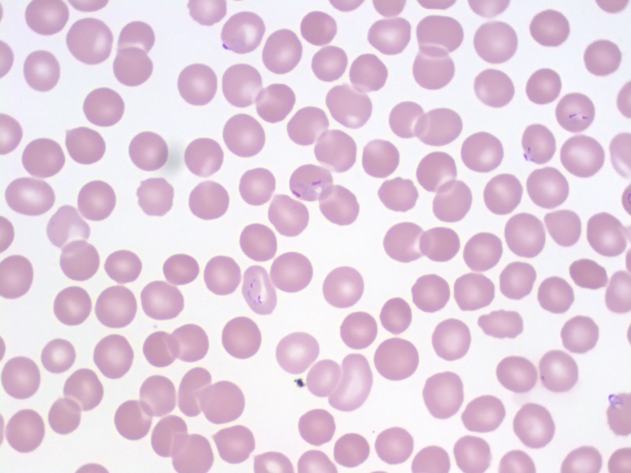
Clinivue software evaluates visual features within captured microscopy imagery to support structured review. This visualization is conceptual and does not expose proprietary scoring.
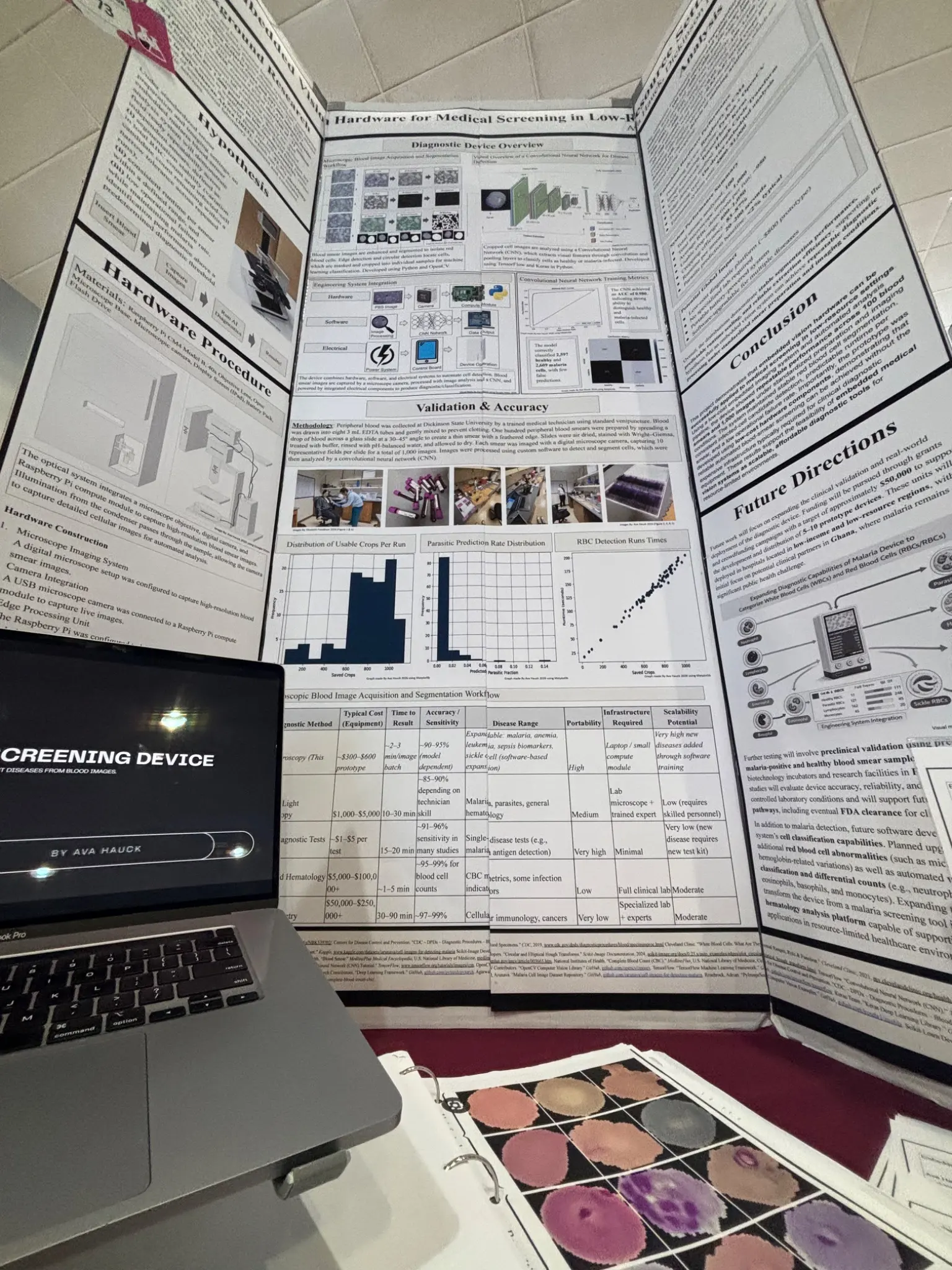 RESEARCH / ITERATION
BUILD. TEST. MEASURE. IMPROVE.
Current research includes malaria-focused laboratory evaluation. Anemia, sepsis, and leukemia modules remain experimental software research and are not laboratory validated.
Explore the research ↗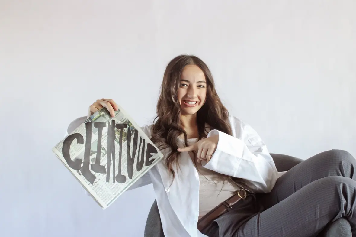
OUR STORY
Before Clinivue was a company, it was a question asked by a student from Dickinson, North Dakota—and the decision to keep building until the question became hardware.
Enter the story →THE NEXT CHAPTER
Clinivue is building toward that future.