Blood smears analyzed
RESEARCH / EVIDENCE
Build. Test. Measure. Improve.
Clinivue's development is being driven by iterative engineering, microscopy research, software development, and laboratory evaluation.
SCROLL TO EXPLORE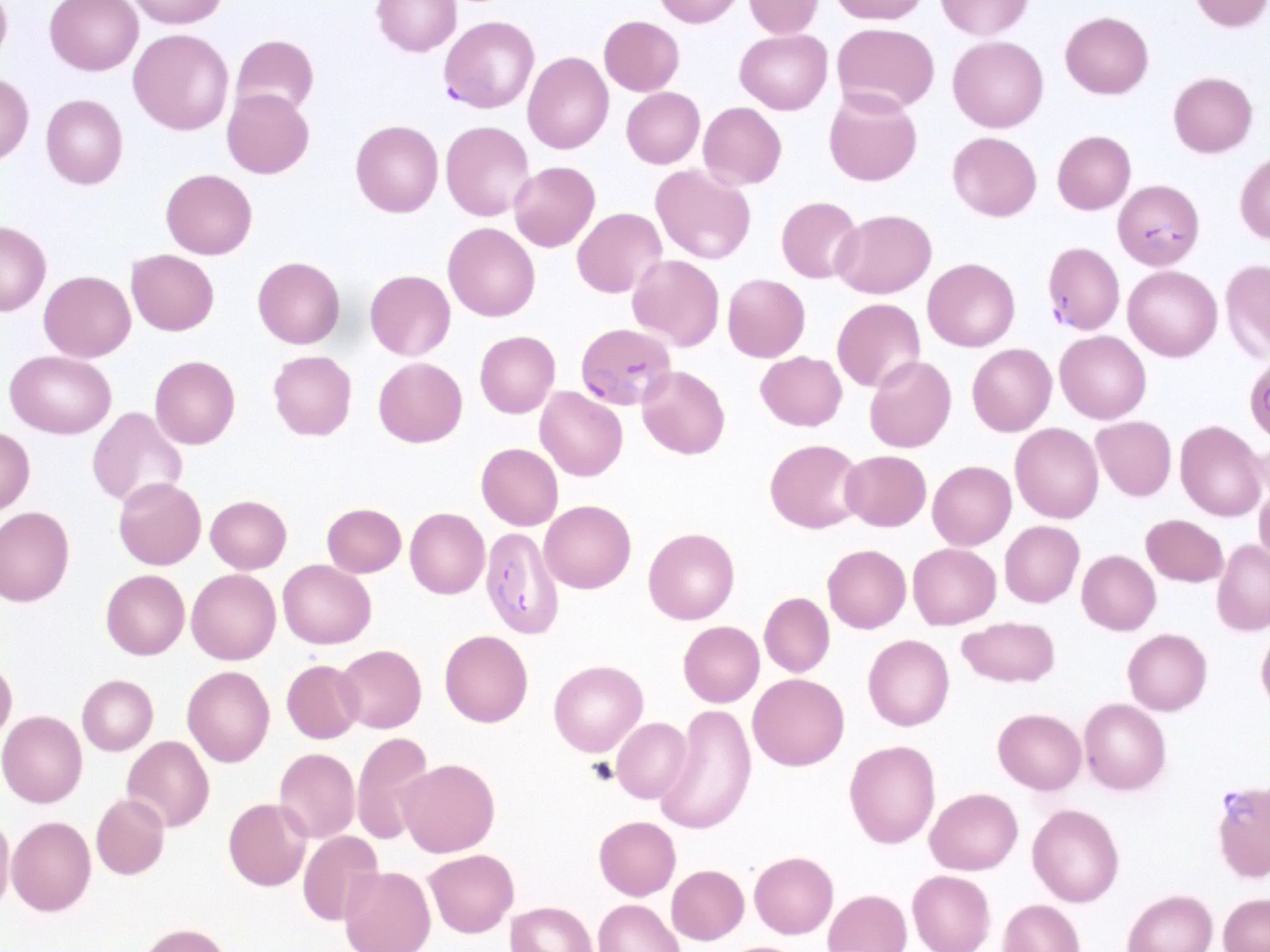
CURRENT RESEARCH
REAL MICROSCOPY → DIGITAL CAPTURE
The research begins in the field of view.
The biological structures in this uploaded malaria-positive microscopy image are unaltered. Restrained markers correspond only to founder-confirmed reference locations and illustrate visual inspection—not Clinivue’s proprietary scoring or a clinical diagnosis.
RESEARCH RESULT
FOUNDER-LED EXPERIMENTAL RESEARCH
From prototype to evidence.
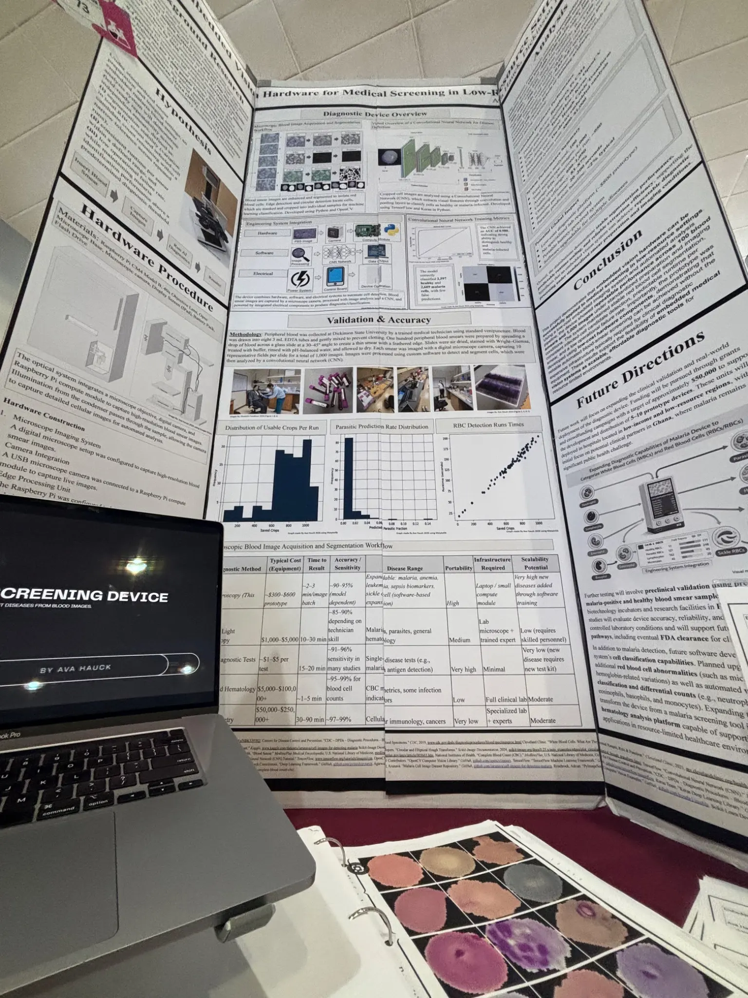
Research prototype validation from Embedded Vision Hardware for Medical Screening in Low-Resource Settings. These results are not peer-reviewed clinical evidence.
Microscopy images analyzed
CNN ROC AUC
Average RBC detections / run
Usable crop range
Cropping efficiency
Prototype processing time
Healthy / malaria-infected cells correctly classified in reported test results
Study Context & Limitations +
Experimental research conducted in a laboratory/prototype setting; not FDA-cleared and not a prospective clinical diagnostic trial.
Performance may change across patient populations, specimen preparation, imaging environments, equipment, operators, and future device configurations. Independent validation is required.
Malaria is the current research hero application. Anemia, sepsis, leukemia, urine, and cervical-cytology work remain software R&D or future platform expansion.
AUC measures discrimination across thresholds; it is not equivalent to 98% clinical accuracy.
EXTERNAL EVIDENCEWhy standardization matters.Explore the clinical context +
There is no single “manual accuracy” number.
Microscopy performance depends on specimen preparation, equipment, reagents, workload, training, quality assurance, reader experience, setting, patient population, and parasite density. Clinivue is being developed to add standardized image acquisition and software-assisted review—not to diminish the role of skilled microscopists.
WHO Malaria Microscopy Quality Assurance Manual ↗88 clinical laboratories
One cross-checking evaluation reported 79.4% sensitivity and 53.8% specificity, with 37.7% fully correct, 25.8% acceptable, and 36.5% incorrect responses against reference review.
Mukadi-Kaningu et al. Performance cannot be generalized to all laboratories, countries, or populations.42.7% / 99.3%
Microscopy sensitivity / specificity versus PCR in the reported study.
Shankar et al. · DOI ↗65.3% / 98.2%
Microscopy sensitivity / specificity versus nested PCR among asymptomatic pregnant women in the reported study.
Adebusuyi et al. · DOI ↗Published performance varies substantially by setting and methodology. These examples do not describe all hospitals or all microscopists and do not establish that Clinivue improves clinician accuracy.
METHODS / SOFTWARE
Microscopy becomes structured review.
A public view of the research workflow without exposing proprietary model architecture, scoring, thresholds, or training methods.
01Prepared smear
02Image capture
03Cell segmentation
04CNN analysis
05Structured review
RAW MICROSCOPYAI-ASSISTED REVIEW
Clinivue software evaluates visual features within captured microscopy imagery to support structured review. This visualization is conceptual and does not expose proprietary scoring.
Malaria
Founder-led laboratory proof of concept.
Anemia · Sepsis · Leukemia
Not laboratory validated.
Urine · Cervical cytology
Future platform expansion.